

Retinopathy & Macular Care
Retinopathy & Macular Care
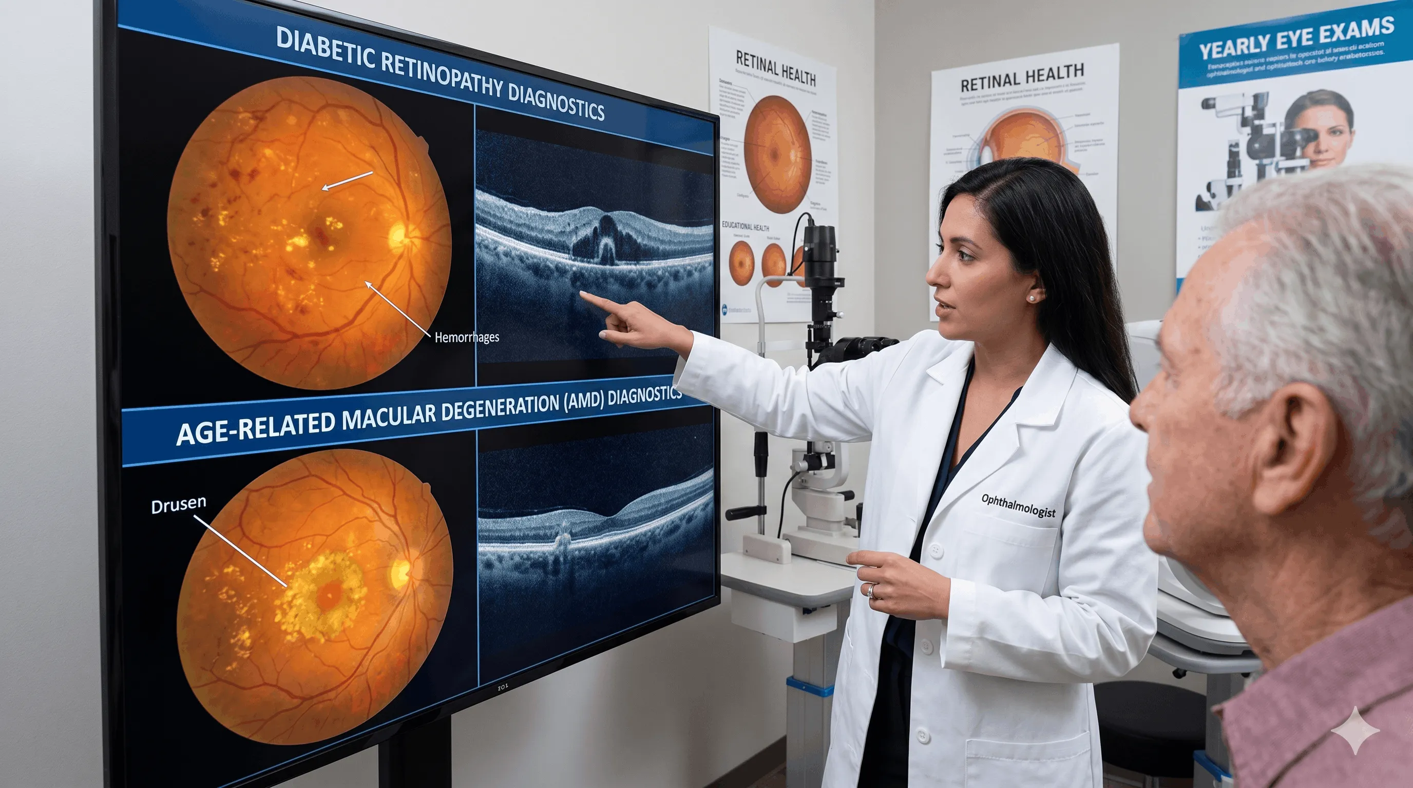
Retinal health is paramount. We specialize in managing Diabetic Retinopathy, where blood vessels are damaged by sugar levels, and Age-Related Macular Degeneration (AMD), which affects central vision. Early detection via yearly exams is critical for specialized treatment.
Our Treatment Benefits
Center-Vision Health
Maintaining the ability to read and recognize faces.
Preventive focus
Screening diabetic patients for early vascular changes.
Our Process
In-Depth Imaging
High-resolution mapping of retinal layers.
Emergency Path
Rapid treatment for sudden vision changes.
Vascular Control
Managing vessel health with targeted enjections.
Patients diagnosed with age-related macular degeneration or diabetic retinopathy requiring ongoing management. Regular ophthalmic screenings are essential to determine the timing and necessity of intervention. Patients must have realistic functional expectations.
Recovery varies significantly by treatment modality, such as intraocular injections or laser therapy. Mild blurred vision or localized irritation is expected immediately after treatment. Normal routines can usually be re-established within a day.
All treatments carry inherent clinical risks. These may include localized bleeding, intraocular infection, or potential loss of vision if the underlying condition aggressively progresses despite optimal intervention.
Maintain strict control of blood sugar levels if diabetic to support overall ocular health. Attend all follow-up appointments diligently, and avoid strenuous physical activity immediately following any clinical procedure.
Frequently Asked Questions
Diabetes cannot only be treated as a blood sugar disease; it is also a significant risk factor affecting the retinal vessels in the eye. Diabetic retinopathy might not show symptoms initially. While the patient thinks they see perfectly well, leakage, edema, bleeding, or the formation of new vessels may have started in the retinal vessels. This situation depends on the development of leakage and circulation disorders in the retinal vessels. For this reason, the 'if there is no complaint, there is no problem' approach is not correct for diabetic eye patients.
In a retinal exam, fundus evaluation is used, alongside OCT and angiographic examinations when necessary. The goal is to detect damage before the vision drops. The risk increases as the duration of diabetes lengthens and blood sugar control deteriorates. Conditions like hypertension, kidney disease, and pregnancy make the follow-up plan even more critical. When the macula is involved, central vision is more significantly affected; hence, simply looking at visual acuity poses a risk.
In treating diabetic retinopathy, blood sugar control is the main element; but it may not always be enough alone. Intraocular injections, laser therapy, or vitrectomy in advanced cases may be necessary. In patients followed up from the early period, the chance of preserving vision is much higher. Thus, my recommendation to diabetic patients is clear: Even if your vision is fine, do not neglect regular retinal checks. Because diabetes is a disease that progresses silently in the eye, but takes a heavy toll when discovered late. A single examination is not considered sufficient in retina tracking; the frequency is affected by the duration of diabetes, HbA1c level, pregnancy status, and accompanying vascular diseases. While a yearly follow-up may suffice for some patients, a more frequent OCT or injection plan is needed for others. The good news is: the chance of preserving vision is high when diabetic retinal disease is caught early. If delayed, treatment turns into a longer and harder process.
Retinal tear and retinal detachment are among the eye conditions requiring urgent evaluation. The most typical symptoms are sudden-onset light flashes, newly occurring or significantly increasing floaters, feeling a curtain or shadow over the field of view, and sudden blurred vision. Especially the expression 'it's as if a black curtain is coming from the side' must be taken very seriously regarding retinal detachment.
These complaints may not always mean a detachment; sometimes it could be more benign processes like vitreous separation. But a patient cannot distinguish this on their own. The detailed retinal exam will make the distinction. The risk is higher in high myopes, after trauma, in those who have had previous cataract surgery, and in those with a family history.
If a retinal tear is caught early, surrounding it with a laser can control the problem before a detachment develops. If retinal detachment has developed, surgery is needed, and time is crucial. Intervening, especially before the macula is involved, is determinative for visual prognosis. Because if the macula is affected in a retinal detachment, even if the retina is anatomically put back in place, functional vision may never return to its former level. Therefore, someone experiencing sudden floaters, light flashes, or a curtain sensation should not say, 'let me wait a bit, it will pass'; they must be evaluated by an ophthalmologist on the same day. The main goal of an urgent visit is to stop the progression before the macula is affected. Thus, a detailed retinal exam with pupil dilation shouldn't be neglected for patients arriving with these complaints. An early diagnosis can sometimes be resolved merely with a short laser procedure, whereas a late presentation might require much more comprehensive surgeries. In retinal diseases, hours and days can truly change the outcome.
Macular degeneration, medically known as age-related macular degeneration (AMD), is a disease that affects the center of the retina and specifically impairs detailed vision. Patients might describe complaints like seeing texts crooked, an inability to recognize faces, or feeling a blurry or empty space in the center. There are dry and wet types; especially in the wet type, early diagnosis becomes much more critical.
Here we need to be realistic: In macular degeneration, the goal is not always to return the eye entirely to its former state. The goal of the treatment is to slow the progression of the disease, protect preserving existing vision, and, if possible, achieve a gain. For the wet type, intraocular injection treatments are among the most important options today. For the dry type, currently, there is no definitive treatment that entirely eliminates the disease; regular follow-up, lifestyle adjustments, and nutritional supplements are evaluated in suitable patients.
Waiting and deciding on your own in macular diseases is risky. Because some patients realize the loss in the other eye late since they see well with one eye. The disease may not affect both eyes simultaneously and at the same speed. Amsler grid follow-up, OCT examination, and regular retina control are essential. Especially for people over age 50, smokers, and those with a family history, even the slightest change in central vision should be taken seriously. One of the factors determining treatment success in macular disease is the patient's follow-up discipline. Skipping appointments, especially in the wet type requiring injection therapy, can lead to the loss of gained vision. Quitting smoking, protecting vascular health, and regular checks are just as valuable as medicine for this disease. People noticing breaking, bending, or shifting of letters in central vision should attend a retina evaluation without waiting.
The complaint of eye floaters is frequently related to natural changes in the vitreous gel and does not always mean a bad scenario. However, sudden onset, rapidly increasing floaters; accompanying light flashes; and especially a sensation of a shadow or curtain in the visual field must be taken seriously. Because these symptoms can be heralds of a retinal tear or detachment.
The biggest mistake patients make is reading about these complaints on the internet and interpreting them on their own. Only a detailed eye fundus examination will show whether the floater is benign or risky for the retina. Highly myopic individuals, those having experienced trauma, and patients post-cataract surgery should be particularly cautious.
Even if no problem is detected during the exam, a short-term re-check may be recommended for some patients. Because small tears not seen in the first exam can become pronounced in the following days. In summary: A few old, static floaters are generally less concerning; but new onset or rapidly multiplying floaters must never be underestimated. The most expensive mistake in eye health is assuming an urgent sign is normal. A scenario alarming for retinal risk is the shape-shifting of the complaint. A few floaters that have stayed the same for years and a suddenly increasing floater accompanied by flashes from last night are not the same thing. Especially signs suddenly intensifying in one eye should be evaluated more carefully. Therefore, the patient should know their normal, and come in for an exam without delay when noticing a change.


Experience Premium
Eye Care Today
Take the first step towards clearer vision. Select a convenient date and time for your consultation with our specialist team.
Book Your Consultation
Choose a convenient time for your assessment with Dr. Söyük.